
Boundaries of optical microscopy stretched
on
The field of optical microscopy has developed rapidly in recent years. Through the invention of the so-called super-resolution fluorescence microscopy, it has recently become possible to observe even the smaller parts of a living cell. With a smart refinement to the super-resolution technique, researchers from the Technical University Delft, The Netherlands have stretched the boundaries of what is possible. Using their method it is possible to focus on structures 3 nanometres in size.
Diffraction limit
The diffraction limit is a theoretical limit up to which two adjacent points can still be distinguished with an optical microscope. The diffraction limit is also a function of the wavelength of light. With a conventional optical microscope, according to the theory, the smallest object that can be brought into focus is half that of the wavelength used. Anything that is smaller cannot be brought into sharp focus.
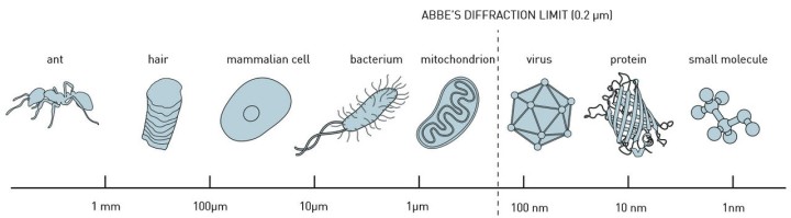
Diffraction limit (image Johan Jarnestad/The Royal Swedish Academy of Sciences).
Fluorescent
The diffraction limit appeared for a long time to be a hard boundary, determined by the laws of nature. But by utilising some clever tricks, physicists nevertheless succeeded in exceeding the theoretical limit. In super-resolution fluoresce microscopy, certain proteins or molecules are made fluorescent by genetic modification. The weak light signal that is emitted can subsequently be detected by an optical microscope. The problem, however, is that in practice not all proteins of a particular type can be labelled this way, but only about 30 to 50 percent. You can then observe a number of individual luminous points, but not the entire structure.
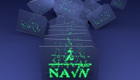
To solve this problem, the Delft researchers have devised a modification to the super-resolution microscopy, which is comparable what is known as ‘compositing’ in photography: the stacking of multiple images to create a compound image. The averaging of the data from the various measurements has already been done in electron microscopy, but that is a completely different technology. It took two years to adapt this technique for optical microscopy.
Graphics card
One problem was also that much computing power is required to combine the data of hundreds, if not thousands, of ‘snapshots’. But thanks to the computer games industry there are now graphics cards available that are very good at parallel computation. The result is that measurements can be combined into a single image in a few hours.
The research brings electron microscopy and optical microscopy closer together. This is important because both techniques give a different view and are therefore complementary, but are still far apart in terms of their capabilities. The best electron microscopes are 30 to 50 times more powerful than the best optical microscopes. Bringing these two worlds closer together could lead to new biological insights.
According to the researchers, their technique, which currently reaches a level of 3 nanometres, should eventually also be able to image structures 1 nanometre in size. The dimensions of the fluorescent labels become a limiting factor.
The findings of the researchers have been published in the journal Nature Methods.
Discussion (0 comments)