
Ultra-thin optical fiber yields super-sharp images
February 3, 2016
on
on
Thanks to collaborative research by scientists at the MESA+ Institute for Nanotechnology of the University of Twente (UT), the Max Planck Institute for the Science of Light (MPL), FOM and Carl Zeiss AG, super-sharp imaging inside the human body with tiny endocopes is a step closer to reality. The combination of unique optical fibers from MPL and advanced wavefront shaping technology from UT allows light to be focused with unprecedented high resolution without using lenses.
Presently the best possible resolution of fiber optic endoscopes is one micron, which is not sharp enough to see the internal details of tissue cells or other tiny features. Some endoscopes consist of a large bundle of glass fibers, with each fiber representing one pixel. These bundles are usually fairly thick – at least 1 mm in diameter. Endoscopes using multimode fibers give a better image and can be as thin as 0.1 mm. With these fibers the resolution is mainly limited by the fact that the fiber only transmits light along its axis. Light that enters at a small angle can still be transmitted by reflection from the fiber walls, but if the angle is too large the light leaks out through the walls. The researchers showed that this limitation can be overcome by using photonic crystal fibers.
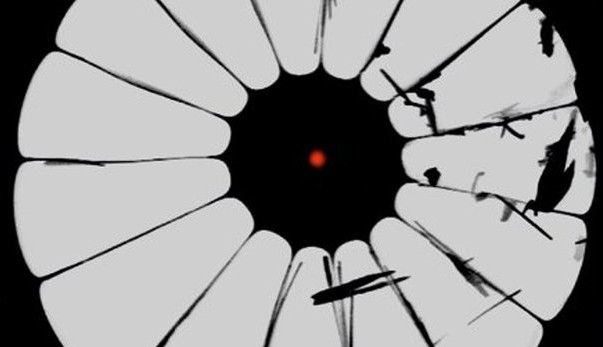
Conventional step-index glass fibers consist of a core and a cladding made from different materials with distinctly different refractive indexes, causing light to propagate along the fiber axis by total internal reflection. Photonic crystal fibers are made from a single material, and the light is guided by a specific pattern of air-filled voids in the cladding. A unique feature of these fibers is that the cladding structure can be engineered to obtain specific optical fiber properties. In this research the scientists designed and built a photonic crystal fiber that can focus a beam of visible red laser light passing through the fiber to a spot 0.52 µm in diameter.
Presently the best possible resolution of fiber optic endoscopes is one micron, which is not sharp enough to see the internal details of tissue cells or other tiny features. Some endoscopes consist of a large bundle of glass fibers, with each fiber representing one pixel. These bundles are usually fairly thick – at least 1 mm in diameter. Endoscopes using multimode fibers give a better image and can be as thin as 0.1 mm. With these fibers the resolution is mainly limited by the fact that the fiber only transmits light along its axis. Light that enters at a small angle can still be transmitted by reflection from the fiber walls, but if the angle is too large the light leaks out through the walls. The researchers showed that this limitation can be overcome by using photonic crystal fibers.
Conventional step-index glass fibers consist of a core and a cladding made from different materials with distinctly different refractive indexes, causing light to propagate along the fiber axis by total internal reflection. Photonic crystal fibers are made from a single material, and the light is guided by a specific pattern of air-filled voids in the cladding. A unique feature of these fibers is that the cladding structure can be engineered to obtain specific optical fiber properties. In this research the scientists designed and built a photonic crystal fiber that can focus a beam of visible red laser light passing through the fiber to a spot 0.52 µm in diameter.
Read full article
Hide full article
Discussion (0 comments)