
Ultra-Thin Endoscope Produces High Quality Images
December 10, 2019
on
on
Under the direction of Mark Foster, a lensless, ultra-miniaturized endoscope has been developed at the John Hopkins University. The new design surpasses conventional micro-endoscopes in image quality and by virtue of its slender cross-section can be used less invasively in medicine. The research results are published in the journal Science Advances.
When conventional endoscopes are scaled down in size the resulting smaller lenses and progressive miniaturization results in poorer quality images. Currently, standard micro-endoscopes have a diameter greater than 0.5 mm and require relatively large lenses in order to produce high quality images. Lensless micro-endoscopes already exist but the optical fibre they use to scan an area of the brain pixel by pixel often bends and loses imaging ability when flexed.
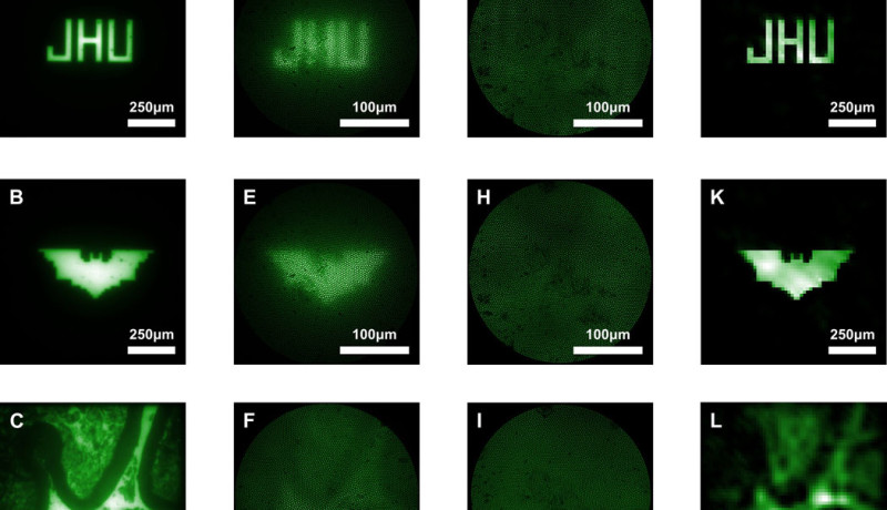
Despite its smaller size, the new lensless, ultra-miniaturized micro-endoscope has none of these disadvantages. According to the JHU press release It uses a coded aperture, or flat grid that randomly blocks light and creates a projection in a known pattern akin to randomly poking a piece of aluminum foil and letting light through all of the small holes. This produces a messy image, but one that provides lots of information about where the light originates, and that information can be computationally reconstructed into a clearer image.
This new design of micro-endoscope does not need to be moved to focus on objects at different distances. It uses computational refocusing to determine from where the light originates in 3 dimensions. These properties are of particular interest in brain surgery and for imaging of neural activity.
When conventional endoscopes are scaled down in size the resulting smaller lenses and progressive miniaturization results in poorer quality images. Currently, standard micro-endoscopes have a diameter greater than 0.5 mm and require relatively large lenses in order to produce high quality images. Lensless micro-endoscopes already exist but the optical fibre they use to scan an area of the brain pixel by pixel often bends and loses imaging ability when flexed.
Despite its smaller size, the new lensless, ultra-miniaturized micro-endoscope has none of these disadvantages. According to the JHU press release It uses a coded aperture, or flat grid that randomly blocks light and creates a projection in a known pattern akin to randomly poking a piece of aluminum foil and letting light through all of the small holes. This produces a messy image, but one that provides lots of information about where the light originates, and that information can be computationally reconstructed into a clearer image.
This new design of micro-endoscope does not need to be moved to focus on objects at different distances. It uses computational refocusing to determine from where the light originates in 3 dimensions. These properties are of particular interest in brain surgery and for imaging of neural activity.
Read full article
Hide full article
Discussion (0 comments)